Arthroscopy of the Knee
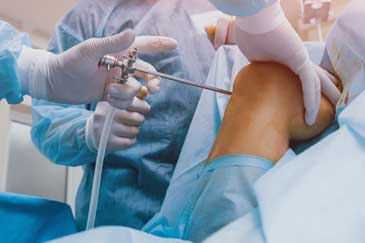
Knee arthroscopy or arthroscopy of the knee is a surgical technique in which we can diagnose or manage and treat many common knee injuries and knee conditions. During the knee arthroscopy procedure, Dr. Hudanich inserts a small camera, called an arthroscope, into your knee joint. The camera displays images on a video monitor, which your orthopedic surgeon uses to guide miniature surgical instruments.
What is Arthroscopy of the Knee?
Dr. Hudanich makes a very small incision to insert a tiny camera into your knee during the knee arthroscopy procedure. This is called an arthroscope. It can also repair the ligaments of the joint. Ligaments are a short band of tough, flexible, fibrous connective tissue that connects two bones or cartilages or holds together a joint.
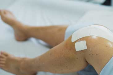
Because the arthroscope and surgical instruments are thin, the incisions made are very small incisions, rather than the larger incisions usually needed for open surgery.  This results in less pain for the patient, less joint stiffness, and often shortens the recovery time. You will be able to start rehabilitation and return to your favorite activities much quicker.
The Anatomy of the Knee
Did you know that your knee is the largest joint in your body and one of the most complex? The bones that make up the knee include the lower end of the thighbone or femur, the upper end of the shinbone or tibia, and the kneecap or patella.
Other important structures that make up the knee joint include:
- Articular cartilage. The ends of the femur and tibia, and the back of the patella are covered with articular cartilage. This slippery substance helps your knee bones glide smoothly across each other as you bend or straighten your leg.
- What is the Synovium? The synovium, also referred to as the synovial membrane, is the soft tissue that lines the spaces of diarthrodial joints, tendon sheaths, and bursae. The synovium is a thin lining of the entire inner surface of your knee joint, except where the joint is lined with the cartilage. This lining releases a fluid that lubricates the cartilage and reduces friction during movement.
- What is the Meniscus? The meniscus is a piece of cartilage in your knee that cushions and stabilizes the joint. It protects the bones from wear and tear. Two wedge-shaped pieces of meniscal cartilage act as shock absorbers between your femur and tibia. Different from articular cartilage, the meniscus is tough and rubbery to help cushion and stabilize the joint.
- What are Ligaments? Our bones are connected to our other bones by ligaments, of which the four main ligaments in your knee act like strong, powerful ropes to hold the bones together and constantly keep your knee stable.
- The two collateral ligaments are found on either side of your knee.
- The two cruciate ligaments are found inside your knee joint. They cross each other to form an "X" with the anterior cruciate ligament in front and the posterior cruciate ligament in back.
Among the most common knee injuries we treat are meniscus tears. A meniscus tear may happen most if you are a professional athlete, like a basketball or football player. You may also tear your meniscus in noncontact sports requiring jumping and cutting, such as skiing, volleyball and soccer, or during activities in or outside the house.
What is a lateral or medial meniscus tear?
Menisci, the medial meniscus and the lateral meniscus, are crescent-shaped bands of thick, rubbery cartilage attached to the shinbone or tibia. They act as shock absorbers and stabilize the knee. The medial meniscus is on the inner side of the knee joint. A meniscal tear is one of the most frequently occurring cartilage injuries of the knee. A simple twist of your knee and you may have torn your meniscus. In some incidents, a piece of the shredded cartilage breaks loose and catches in the knee joint, causing it to lock up.
Tears may happen by falling down, changing direction suddenly while running, and often occur at the same time as other knee injuries, like an anterior cruciate ligament or ACL injury. Meniscus tears are a special risk for older patients since the meniscus weakens with age. More than 40% of people 65 or older have them.
If you have a torn meniscus, you may have heard and felt a popping in your knee.
 You are in pain and your knee swells up.
You may not be able to walk, bend or straighten the leg.
A tendency for your knee to get stuck or lock up.
Depending on how badly you injured your knee, at first the pain may be extremely bad or not bad at all. Once the inflammation sets in, your knee will probably hurt quite a bit.
A medial meniscus tear is more common than a lateral meniscus tear. The medial meniscus is on the inner side of the knee joint. The lateral meniscus is on the outside of the knee. Meniscus tears can vary widely in size and severity.
If you are diagnosed with a lateral meniscus tear, the injury is a disruption of the fibrocartilage, the meniscus, which absorbs shock and lubricates the weight-bearing compartment of the knee. The most common cause is excessive pressure and torsional force applied to the fibrocartilage of the outside, lateral meniscus, aspect of the knee. This usually occurs, often as an unrecognized event, during a twisting or hyperflexion or excessive bending injury to the knee.
Most typical symptoms are pain localized at the outside of your knee, lateral, aspect of the knee usually aggravated by vigorous activities and squatting. Mechanical symptoms of catching, popping or even locking may occur. Discomfort is often noted at night and descending stairs.
What is a cartilage tear?
Your knee cartilage can be injured as the result of a traumatic injury, degenerative arthritis, or chronic overuse. There is a difference in a meniscus tear and a cartilage tear. As the knee joint has two types of cartilage inside the joint. One of the types of cartilage is called articular cartilage. The articular cartilage forms the smooth layer of the joint that covers the bone ends. A layer of articular cartilage covers the end of the thigh bone, the top of the shin bone, and the back of the kneecap. The meniscus is a different type of cartilage that forms a shock absorber between the bones. The meniscus is not attached to the bone like the articular cartilage, but rather sits between the bone ends to cushion the joint.
Depending on the type of injury, the different types of cartilage may be damaged. When cartilage is damaged, often it is described as a tear of the cartilage. Typically, when someone refers to a tear in the cartilage, they are talking about an injury to the meniscus cartilage.
It may take up to six weeks for the knee joint to re-establish normal joint fluid after arthroscopic surgery. You may not realize the full benefits of your arthroscopic knee surgery for four to six weeks.
Additional helpful information provided by the American Academy of Orthopaedic Surgeons

